Ultrasonography
We are here for your care.
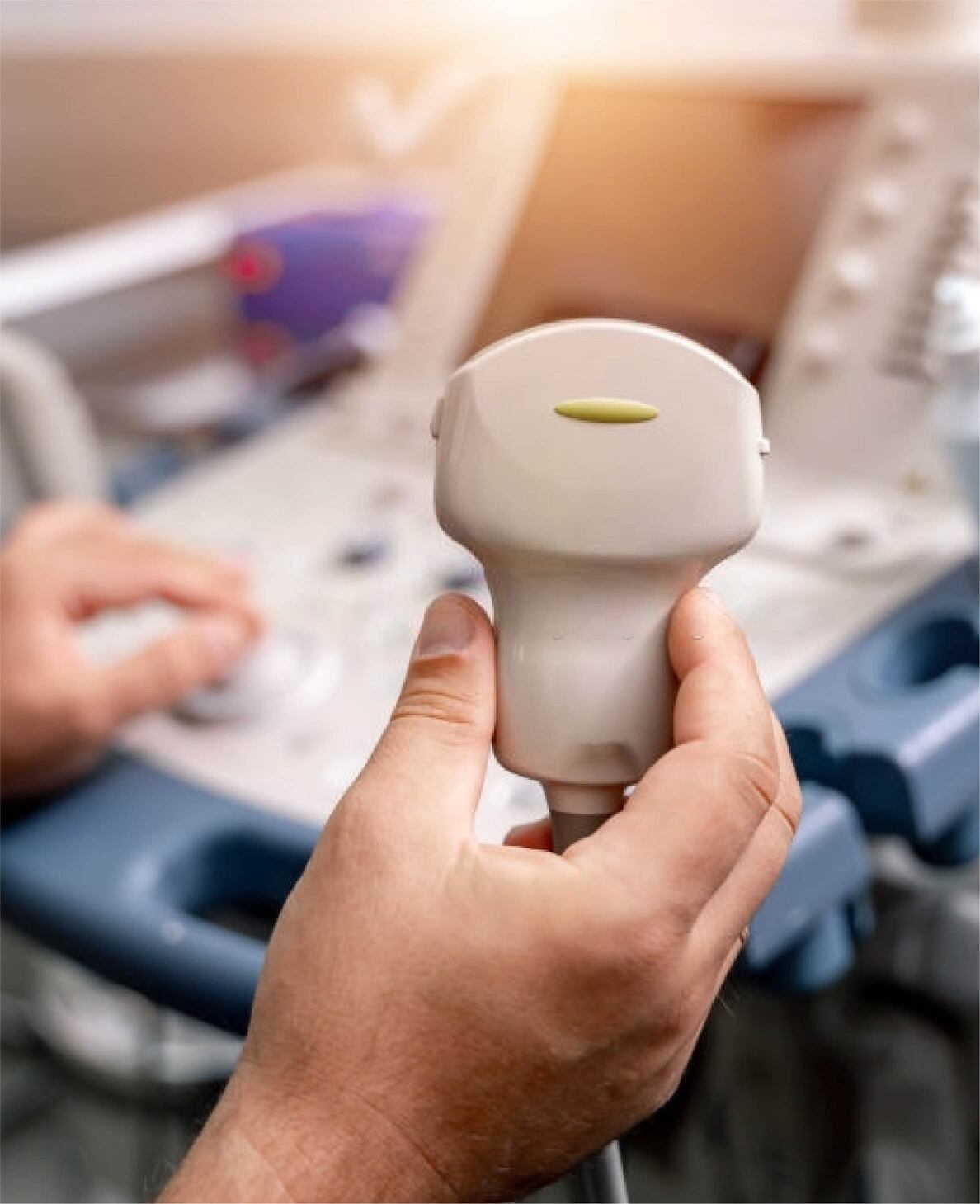
Ultrasonography uses high-frequency sound waves to assess the status of internal organs and tissues. It produces black and white images. Sound waves of 1-10 million hertz are sent through a transducer which is placed over body structures. There are crystals at the top of the transducer that the sound waves bounce back to. Sound waves can be absorbed too.
The regions, that contain fluid or are hollow, such as vessels and bladder, let sound waves pass through them and look black on the screen. Regions that have tissues don’t let the sound waves pass fully, allow refraction of sound, and look grayish white on the screen. The structures that are very hard, like bone, do not let sound waves pass through them at all and, as a result, the sound waves bounce back to the transducer. Such structures appear bright white.

Uses of ultrasonography
The images obtained from ultrasonography are taken rapidly enough to display the motion of organs and structures in the body in real time.
It is used to detect growths and foreign objects that are situated near to the surface of body. It is also used to image internal organs in the abdomen, chest, and pelvis. Moreover, ultrasonography can be used to guide doctors when they are taking a sample of tissue for biopsy. It shows both the position of the biopsy instrument and the area that the biopsy is to be performed on, thus allowing the doctors to see where to insert the instrument and guiding the instrument to its target.
There are three modes to display the information obtained from ultrasonography – A mode, B mode and M mode. In A mode, information is displayed in the form of spikes on a graph. In B mode, the information is displayed in the form of a 2-dimensional anatomical images. M-mode is used to display information as continuous waves to show moving structures. The B-mode is the most common way of using ultrasonography.
For pregnant women, ultrasonography is done to obtain an image of the womb. On the screen, the amniotic fluid will look black, thus allowing the bones and tissues of the baby to be visible (they appear white). Ultrasonography is usually done to assess the development of the baby, find out the gender and detect abnormalities, if any.
Ultrasonography is used to assess the following:
- • Liver, pancreas, and spleen
- • Blood vessels
- • Urinary tract
- • Female reproductive organs
- • Pregnancy
- • Heart
- • Gallbladder and biliary tract

Prasad Hospital is a multi-speciality hospital located in Brahmpura, Muzaffarpur. Backed by a team of experienced doctors, Prasad Hospital is known for its dedication to provide affordable healthcare.
Address
Prasad Hospital, Brahmpura, Muzaffarpur, Bihar-842003
prasadhospitalmuzaffarpur@gmail.com
Phone
9570996625 / 9570996640
